X-Rays
| Overview | Medical X-rays | Advantages | Disadvantages | Uses Medical X-rays |
1. Overview:
X-ray is a high-energy electromagnetic radiation. In many languages, it is known as Rontgen radiation, after the German scientist Wilhelm Conrad Rontgen, who represented it in 1895 and named it X-radiation to mean an unidentified type of radiation.
X-ray wavelengths of X-ray are shorter than ultraviolet rays and longer than gamma rays. There is no universally established, severe meaning of the bounds of the X-ray band. Generally, X-rays have a wavelength varying from 10 nanometers to 10 picometers, equivalent to frequencies in the range of 30 petahertz to 30 exahertz.
X-rays can go through many solid substances such as building materials and living tissue. X-ray radiography is commonly used in medical diagnostics such as checking for broken bones, and substance science. However X-rays are ionizing radiation and disclosure to high intensities can be dangerous to health, causing DNA damage, cancer, and a high number of burns and radiation diseases. Their creation and application are strictly limited by public health authorities.
What are medical x-rays?
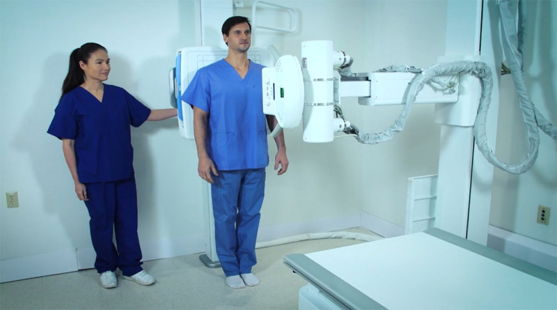
X-rays are a type of electromagnetic radiation, like visible light. Unlike light, however, x-rays have higher energy radiation and can pass through the most substance, including the body. Medical X-rays are used to produce images of tissues and compositions inside the body. If X-rays passing through the body also travel through an X-ray detector on the other side of the patient, an image will be created that signifies the “shadows” formed by the items inside of the body.
One kind of X-ray detector is a photographic background screen, but more other types of detectors are used to generate digital images. The X-ray images that result from this method are named radiographs.
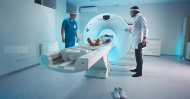
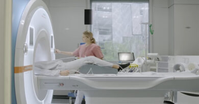
Medical x-rays work:

To create a radiograph, a patient is placed so that the part of the body being imaged is positioned between an x-ray source and an x-ray detector. When the machine is switched on, x-rays pass through the body and are absorbed in special quantities by different tissues, depending on the radiological mass of the tissues they pass through. Radiological density is established by both the density and the atomic number of the objects being imaged. For example, our bones hold calcium, which has a higher atomic number than other tissues. Because of this property, bones readily attract x-rays and therefore generate high contrast on the x-ray detector. As a result, bony structures come into view whiter than other tissues alongside the black background of a radiograph. On the other hand, x-rays pass more easily through less radiological thick tissues, like fat, muscle, and lungs. These structures are shown in shades of gray on a radiograph.
2. Advantages of X-rays:
- Diagnostic imaging: X-rays are frequently used to diagnose a range of illnesses in medical settings. In order to find fractures, tumors, infections, and other abnormalities, they give precise images of the bones, joints, and organs.
- Quick and non-invasive: X-ray procedures typically take little time and cause no discomfort. They are a practical choice for diagnostic imaging because they don’t require any incisions or surgery.
- Economical: X-rays are more reasonably priced than other imaging methods like CT scans or MRIs. As a result, many patients can access them more easily.
- Wide availability: The majority of hospitals, clinics, and radiology centers have access to X-ray machines, making it easy to perform X-rays in these settings.
3. Disadvantages of X-rays:
- Radiation exposure: Ionizing radiation, which can be dangerous in large amounts, is exposed during X-ray imaging. Even though the radiation dose from diagnostic X-rays is ordinarily minimal, prolonged exposure or high levels could be dangerous.
- Limited ability to see soft tissues, such as muscles, tendons, and organs. X-rays are best useful for capturing images of dense structures, such as bones. On X-ray scans, subtle anomalies in these regions might be harder to see.
- Possibility for mistakes and incorrect interpretation: X-ray image interpretation requires a qualified radiologist or other healthcare expert. There is still a chance for mistakes or misunderstandings, which could result in inaccurate diagnoses or the failure to detect anomalies.
- Limited real-time guidance during procedures: Because X-rays produce a static image, using them as real-time guidance during surgical or interventional treatments is difficult. For these uses, other imaging modalities like fluoroscopy or ultrasonography are frequently preferred.
It’s vital to keep in mind that, despite some drawbacks, X-rays are generally considered to be safer than they are beneficial for identifying a variety of diseases. Based on your unique circumstances, your healthcare professional will weigh the potential risks and advantages before recommending the best imaging procedure.
Uses of medical X-rays:
There are some most important uses of medical X-rays:
Diagnostic:
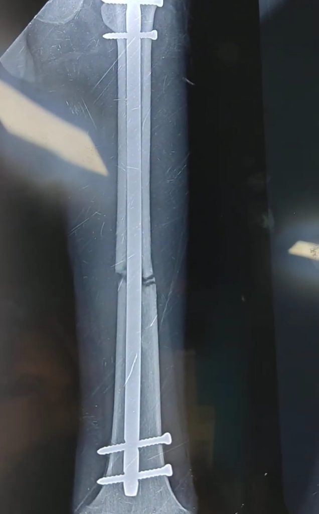
X-ray radiography: Identify bone breakage, certain tumors, and other unusual masses, pneumonia, some kinds of injuries, calcifications, unknown objects, or dental problems.

Mammography: A radiograph of the breast that is utilized for cancer detection and diagnosis. Tumors tend to show regular or irregular-shaped masses that are rather brighter than the background on the radiograph. Mammograms can also detect small bits of calcium, called microcalcifications, which appear as very bright dots on a mammogram. While generally kindly, specific samples of microcalcifications could designate and identify the cancer.
Computed tomography (CT): Combines traditional X-ray technology with computer processing to produce a sequence of cross-sectional images of the body that can later be joined to create a three-dimensional X-ray image. CT images are more complete than simple radiographs and give doctors the facility to examine structures within the body from many different angles.
Fluoroscopy: Uses X-rays and a fluorescent display to take real-time images of movement within the body or to analyze diagnostic processes, such as following the path of an injected or swallowed contrast representative. For example, fluoroscopy is used to observe the movement of the beating heart, and, with the help of radiographic different agents, to analyze blood flow to the heart muscle as well as throughout the blood vessels and organs. This technology is also applied with a radiographic contrast agent to direct an inside the threaded catheter through the cardiac angioplasty, which is a simply invasive procedure for opening the closed blood vessels that supply blood to the heart.
Therapeutic:
Radiation therapy in cancer treatment: X-rays and other types of high-energy radiation can be used to destroy cancer cells by damaging their DNA. The radiation quantity used for treating cancer is much higher than the radiation quantity used for analytical imaging for medical treatment. Therapeutic radiation can come from a device outside of the body or from radioactive objects that are located in the body, inside or near cancer cells, or inserted into the bloodstream.